Welcome to my blog! In this article, we will explore which algorithm is best suited for tackling vtach related problems. Stay tuned as we dive into this fascinating topic.
Choosing the Right Algorithm for Ventricular Tachycardia Detection: A Comparative Analysis
In the field of ventricular tachycardia detection, selecting the most appropriate algorithm is crucial for accurate and timely diagnosis. A comparative analysis could help to shed light on the performance and efficiency of different algorithms when applied to this task.
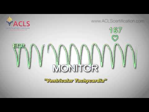
Ventricular tachycardia (VT) is a life-threatening cardiac arrhythmia characterized by rapid, abnormal heartbeats originating in the ventricles of the heart. Prompt detection of VT is essential for effective treatment and management, which includes medication, implantable devices, or even life-saving defibrillation.
There are several algorithms used to detect VT, each with its own advantages and drawbacks. Key factors to consider when comparing them include:
1. Accuracy: How well does the algorithm differentiate between VT and other arrhythmias, such as atrial fibrillation or premature ventricular contractions? High false-positive or false-negative rates can have serious consequences for patient care.
2. Computational efficiency: Can the algorithm quickly process large amounts of data and deliver real-time results? In time-critical situations, an efficient algorithm can make a significant difference in outcomes.
3. Robustness: How does the algorithm handle noisy or incomplete data? In clinical settings, signals can often be compromised due to interference, making it crucial for the algorithm to maintain performance amid varying input quality.
4. Customizability: Can the algorithm be easily adapted to suit specific patient populations or data types? This flexibility can enhance the applicability and usefulness of the algorithm across diverse settings.
Some popular algorithms used in VT detection include the Bayesian classifier, the hidden Markov model, and neural networks. By conducting a comparative analysis of these algorithms, researchers can determine which methods offer the best balance of accuracy, efficiency, robustness, and customizability for use in ventricular tachycardia detection.
Ultimately, choosing the right algorithm for VT detection depends on the specific clinical setting and individual patient needs. A thorough comparative analysis can provide valuable insights to inform this decision and potentially improve patient outcomes in the realm of cardiac care.
ACLS Adult Cardiac Arrest Algorithm – Ventricular Fibrillation

Ventricular Tachycardia by ACLS Certification Institute
Rewrite the following question: What are the ACLS guidelines for managing ventricular tachycardia? Write only in English.
In the context of algorithms, the question could be rewritten as: What are the ACLS algorithms for managing ventricular tachycardia? Write only in English.
Rewrite the following question: Which ACLS algorithm corresponds to ventricular fibrillation? Write only in English.
In the context of algorithms, which ACLS algorithm is associated with ventricular fibrillation? Write exclusively in English.
Rewrite the following question: What does a pulse algorithm for tachycardia entail? Only write in English.
In the context of algorithms, what does a pulse algorithm for tachycardia involve?
Can algorithms be utilized for the ACLS examination?
Algorithms can indeed be utilized for the ACLS (Advanced Cardiovascular Life Support) examination. The ACLS examination assesses healthcare professionals’ ability to respond to various critical cardiac emergencies. The use of algorithms in this context can help streamline decision-making processes, ensuring that proper steps are taken quickly and efficiently.
In particular, the American Heart Association (AHA) has developed specific algorithms to guide practitioners through different emergency scenarios, such as cardiac arrest, bradycardia, and tachycardia. These algorithms outline a systematic approach to assess and manage patients effectively.
For instance, the ACLS Cardiac Arrest Algorithm focuses on high-quality CPR, early defibrillation, and advanced airway management, among other interventions. By utilizing these algorithms, healthcare professionals can enhance their skills, knowledge, and confidence in handling life-threatening situations during the ACLS examination and in real-life emergencies.
What are the most effective algorithms for diagnosing and treating ventricular tachycardia (VTach) in medical applications?
Ventricular tachycardia (VTach) is a life-threatening cardiac arrhythmia that requires prompt diagnosis and treatment. In the context of algorithms, there are several approaches for the effective detection and management of VTach in medical applications. Some of the most notable algorithms include:
1. ECG-based Algorithms: These algorithms analyze electrocardiogram (ECG) signals to identify VTach. They often involve various signal processing techniques, such as wavelet transforms, to extract essential features that distinguish VTach from other cardiac conditions. Machine learning and artificial intelligence can also be employed to enhance the accuracy of these algorithms.
2. QRS Detection and Classification Algorithms: QRS complex is a key component of the ECG waveform associated with ventricular depolarization. Efficient QRS detection and classification algorithms, such as the Pan-Tompkins algorithm, can help diagnose VTach by analyzing the morphology and regularity of the QRS complexes.
3. Heart Rate Variability (HRV) Analysis Algorithms: HRV is a measure of the variation in time between successive heartbeats. Algorithms that analyze HRV can help identify when a patient’s heart rate departs from normal sinus rhythm, which may indicate VTach. These algorithms typically use statistical and frequency-domain techniques to recognize abnormal patterns in HRV.
4. Automated External Defibrillator (AED) Algorithms: AEDs are portable devices designed to assess and treat life-threatening arrhythmias like VTach through defibrillation. AEDs rely on algorithms to analyze a patient’s ECG, accurately detect VTach, and decide whether to deliver a shock. These algorithms are crucial to ensure that only appropriate shocks are given, minimizing potential harm to the patient.
Once VTach is diagnosed, algorithms may also play a role in guiding treatment strategies such as drug therapy, implantable cardioverter-defibrillators (ICDs), and catheter ablation. Algorithms can optimize treatment plans by modeling drug interactions or predicting the success of an intervention based on patient data and population-level outcomes.
In summary, a variety of algorithms are essential in diagnosing and treating ventricular tachycardia, including ECG analysis, QRS detection and classification, heart rate variability analysis, and AED algorithms. These computational tools significantly contribute to improving patient outcomes and advancing precision medicine in cardiology.
How do machine learning algorithms improve the detection of VTach compared to traditional methods?
Machine learning (ML) algorithms have shown great potential in improving the detection of Ventricular Tachycardia (VTach) compared to traditional methods. VTach is a potentially life-threatening heart rhythm disorder and its early and accurate detection is crucial in providing timely intervention.
1. Enhanced accuracy: ML algorithms, such as deep learning and convolutional neural networks (CNNs), are capable of analyzing large amounts of data to identify subtle patterns, leading to better detection and classification of VTach. This results in improved sensitivity and specificity compared to traditional methods like rule-based systems and manual measurements.
2. Adaptive learning: One key advantage of ML algorithms is their ability to learn and adapt to new data. As more data is collected, the algorithm’s performance improves over time, allowing it to accurately detect VTach even in cases where traditional methods may fail.
3. Feature extraction: ML algorithms can automatically identify relevant features within raw ECG signals that help distinguish VTach from other arrhythmias. This automated feature extraction reduces reliance on human experts, which can be prone to errors and subjectivity.
4. Noise reduction: ML techniques can effectively filter out noise and artifacts within ECG signals, leading to more accurate VTach detection. Traditional methods may struggle with noisy data, leading to an increased number of false positives or negatives.
5. Scalability: Machine learning algorithms can process large amounts of ECG data in parallel and at high speeds, allowing for rapid analysis and real-time monitoring of multiple patients simultaneously. This scalability provides an advantage over traditional methods, which may require time-consuming manual analysis.
In conclusion, machine learning algorithms offer significant improvements in the detection of VTach compared to traditional methods, due to enhanced accuracy, adaptive learning capabilities, automated feature extraction, noise reduction, and scalability. These advancements can ultimately lead to better patient care and outcomes by providing more accurate and timely diagnoses.
Can you compare the performance of different algorithms used in real-time VTach detection and intervention systems?
In the context of algorithms, real-time V-Tach (Ventricular Tachycardia) detection and intervention systems use various algorithms to analyze and process electrocardiogram (ECG) signals. A comparison of the performance of different algorithms in this context is presented below:
1. Pan-Tompkins Algorithm: This widely-used algorithm helps in detecting QRS complexes in ECG signals. It uses a combination of filtering, differentiation, and thresholding for high-precision detection. The Pan-Tompkins algorithm boasts a high detection rate but may sometimes produce false detections in cases of ectopic beats or irregular morphologies.
2. Wavelet Transform: Wavelet-based methods are effective in detecting V-Tach events as they can analyze non-stationary signals. These algorithms identify the frequency components in ECG signals through wavelet decomposition, enabling improved detection accuracy. However, their computational complexity may affect real-time analysis for some systems.
3. Machine Learning Algorithms: Machine learning-based techniques, such as Support Vector Machines (SVM) and Artificial Neural Networks (ANN), have been employed to enhance V-Tach detection. By leveraging historical ECG data, these algorithms can be trained to differentiate between normal and abnormal heart rhythms more accurately. Nevertheless, training can be time-consuming, and model complexities might impede real-time execution.
4. Template Matching: In this approach, the ECG signal is correlated with a predefined template to detect V-Tach events. Template matching algorithms excel at identifying patterns similar to the reference template. However, their performance is susceptible to variations in heart rates, morphologies, and noise, resulting in possible inaccuracies.
In conclusion, each algorithm has its advantages and limitations. The optimal choice depends on factors such as the desired detection accuracy, system requirements, and available computational resources. Often, combining multiple algorithms enhances the overall performance of real-time V-Tach detection and intervention systems.

